Tag: cosmetic

  • About ‘bags’ under the eyes and cosmetic eyelid surgery

    Tired look, Dr Dion Paridaens explains the surgical options..

    In the cosmetic eyelid surgery practice of the ELZA clinic in Zurich and the Focusclinic of the Rotterdam Eye Hospital I see many clients who are keen to have an upper or lower eyelid correction. Most have a mild to moderate degree of skin excess, some even severe. Smokers often present themselves with festoons: a skin fold, sometimes filled with moisture, which hangs like a kind of bag at the bottom of the lower eyelids. Eyelids may also exhibit excess fat, especially noticeable in overweight or well-known dermatochalazis (skin excess due to loss of elastin fibers). Bags under the eyes or skin excess in the upper eyelids based on dermatochalazis contribute to a tired expression, which often does not reflect the energy status of the client.

    Eyelid correction for bags under the eyes versus upper eyelid correction

    Lower blepharoplasty (eyelid correction) is more complex from a surgical point of view and is less forgiving than upper eyelid correction, which is a very common procedure. Starting and unexperienced surgeons, especially those who have not had proper training in this field of cosmetic surgery through a fellowship program, will soon find out that cosmetic lower eyelid surgery is rather complex, sometimes – unfortunately – at the expense of a good result for their clients.
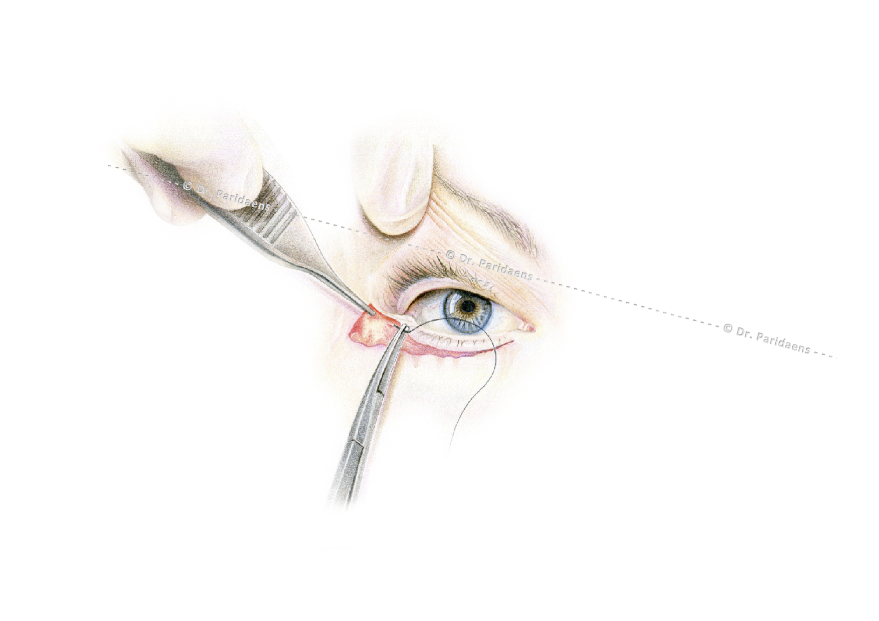
    During lower eyelid correction with skin removal, canthopexy is an essential step: the eyelid is fixed with one or two (buried) stitches on the deeper tissues in the lateral eyelid angle to prevent a postoperative misalignment (eyelid retraction) causing an unnatural and undesirable appearance of whites of the eye under the iris. (Copyright Dr. D Paridaens, oculoplastic surgeon)

    Why is lower eyelid correction more difficult than upper eyelid correction? The importance of canthopexy or canthoplasty in eyelid surgery!

    First, the incisions in the upper eyelids are hidden in the natural crease of the upper eyelid. Only very close to the nose and to the side we can see a temporarily visible scar, which generally heals very well in the month after the operation and can be camouflaged in the initial phase with a foundation creme. In the lower eyelids the scars can be visible for weeks to months, the latter especially in smokers, despite the fact that they are placed in the relaxed skin tension lines (the natural skin lines that occur during the aging process) (this creates “hidden” as it were incisions “).
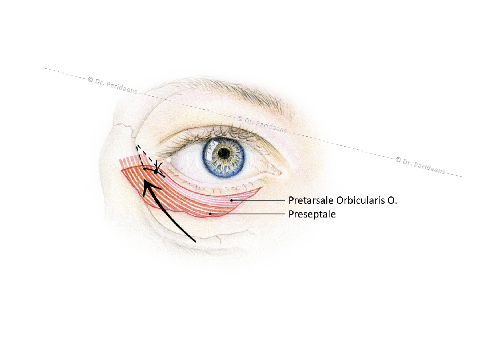
    Fixation of the lower eyelid to the bone membrane with buried sutures and also deep sutures outside the lateral eyelid angle gives a good and natural result. As a result, the lower eyelid is neatly draped around the eyeball in a direction outwards and upwards (see arrow in the figure) and the gravity is neutralized. If these steps are not performed, this can lead to postoperative complications: incorrect position and unsightly retraction of the lower eyelids as well as dry eyes. (Copyright: Dr. D Paridaens, oculoplastic surgeon Illustration: Jessica Leenen)

    Secondly, laxity of the lower eyelid may occur due to weakening and aging of the eyelid tissue and the ligaments that connect the eyelid to the bone. In addition, gravity pulls down the eyelid tissues. If the lateral part of the lower eyelid is not sufficiently shortened or fixed to the bone, any removal of excess skin can lead to an incorrect position of the eyelid (retraction or retraction of the lower eyelid), which can be enhanced by gravity. Therefore, the surgeon should try to prevent this by correcting the lower eyelids by means of 1. adequate canthopexy or canthoplasty (procedures to fix the eyelid to the bone) 2. limiting the amount of vertical skin removal during blepharoplasty, especially when the eye is relatively protruding (proptosis) or when the cheekbone is flat and 3. introduction of an outward / upward vector (force line) that counteracts the gravity vector by placing deeply supporting sutures in the soft tissues just outside the lateral corner of the eyelids.

    Loeb-Hamra technique for eyelid correction: fat redistribution instead of removal!

    If there is too much fat in the lower eyelids while the tear trough or groove between the lower eyelid and the cheek is deep due to tissue loss resulting from aging (which enhances the effect of rings around the eyes), a fat redistribution is more logical than the alternative of removing it. This technique of re-using excess fat to a nearby location with a shortage of fat was described by Dr Loeb and also by Dr. Hamra. Here, after opening the lower eyelid through an opening under the lash line, an opening is made in the orbital septum, a thin connective tissue layer, whereby the fat can bulge forward. Then this fat can be put into a pocket above the orbital rim and placed under or above the periosteum layer above the cheek bone for filling of the tear trough. See the illustrations below.
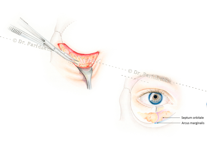
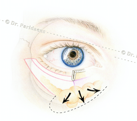
    After the opening of the lower eyelid, the orbital septum is visible behind which the fat excess is located. Near the orbital rim (arcus marginalis) the septum is opened which allows the fat to protrude. (Copyright Dr. Dion Paridaens, oculoplastic surgeon Illustration: Jessica Leenen)
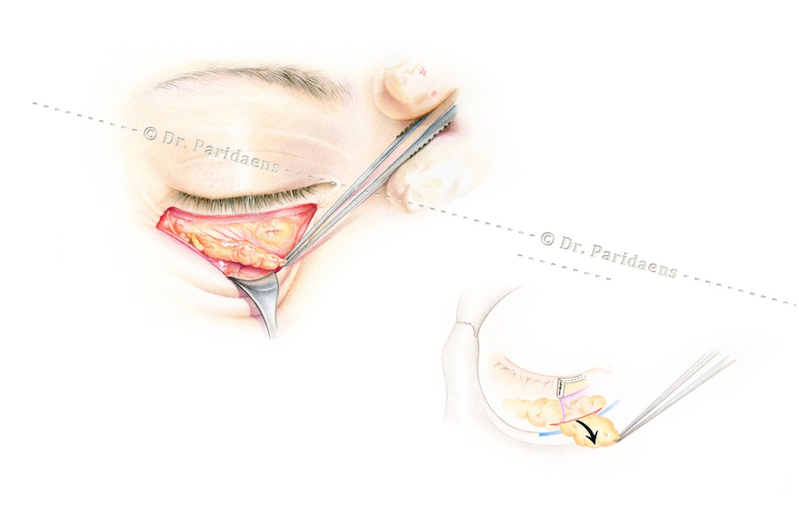
    After the opening of the orbital septum, the surplus of fat – which causes the lower eyelid swelling – can be pulled through the opening and positioned over the orbital rim and put in a pocket above (or under) the periosteum layer above the cheek bone. As a result,  the hollow groove is filled up while the fat acts as a natural filler. (Copyright: Dr. Dion Paridaens, oculoplastic surgeon Illustration: Jessica Leenen)
    The excess of fat can be draped over the lower orbital rim in the direction necessary to fill the hollow groove below  the lower eyelid. (Copyright: Dr. Dion Paridaens, oculoplastic surgeon Illustration: Jessica Leenen)
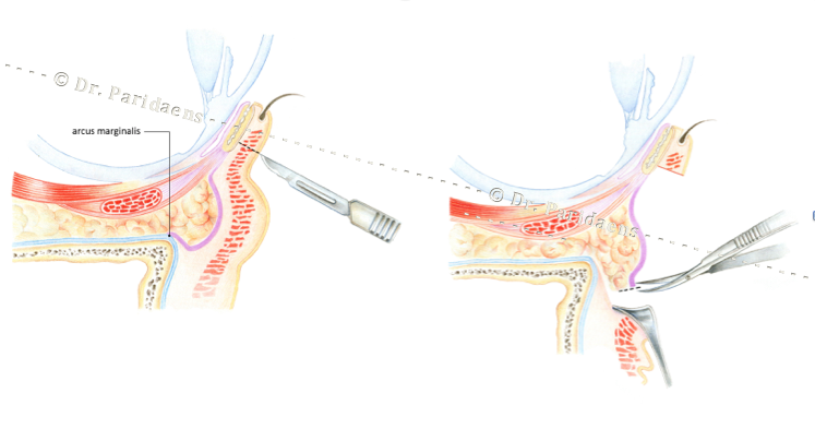
    Left: the lower eyelid skin is incised at just below of the lash line. The skin and Orbicularis Muscle layer is pulled down so that the connective tissue (septum, purple) and the fat excess is visible. Right: the septum is opened at the level of the arcus marginalis, after which the fat can be draped downwards with a decrease of the eyelid swelling as a result. (Copyright: Dr. Dion Paridaens, oculoplastic surgeon Illustration: Jessica Leenen)
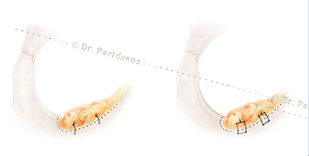
    The excess of fat in the lower eyelid is used for filling the groove under the lower eyelid and fixed to the deep layer with single or mattress sutures. (Copyright: Dr. Dion Paridaens, oculoplastic surgeon Illustration: Jessica Leenen)
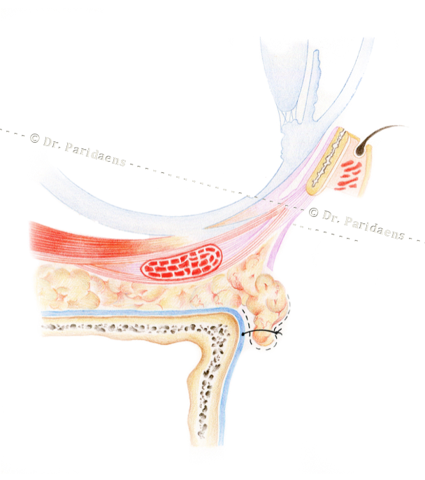
    The fat surplus is draped over the eye socket. As a result, the hollow quarry is filled in, so there is less relief in the lower eyelid / cheek complex with improvement of the cosmetic (Copyright: Dr. Dion Paridaens, oculoplastic surgeon Illustration: Jessica Leenen)

    Additional treatment after eyelid correction with laser or trichloroacetic acid (TCA peeling)

    A well-executed lower eyelid correction generally gives a notable improvement in the appearance because of the reduction of the obvious swelling or excess of skin. However, one cannot expect that the aged skin will recover its original elasticity. Therefore, mild wrinkling or even mild surplus of skin may persist. Additional excision surgery can be hazardous as it might cause unsightly lower eyelid retraction, which may also cause functional problems such as tearing, diminished visual acuity or redness of the eyes.  If the skin is wrinkled, it may be considered to treat the skin with laser or with trichloroacetic acid (TCA peeling) for an improvement of the cosmetic image. In the case of minor abnormalities, laser surgery or peeling can also be chosen instead of surgery.

    Realistic expectations with regard to outcome of cosmetic lower blepharoplasty

    In both upper and lower blepharoplasty it is important that clients are aware about the benefits, side effects and risks of the surgery. Adequate information prior to the surgery should prevent false expectations with regard to the outcome of surgery. If the expectations are not realistic, the outcome may be disappointing for the client even when this is objectively good. Fortunately, in our practice, the vast majority of well-informed and selected clients who have undergone a proper ophthalmologist correction are very satisfied with the result.


    Dr Paridaens is an experienced eyelid and orbital surgeon. He is the current President-Elect of the European Society of Ophthalmic Plastic & Reconstructive Surgery (ESOPRS). In 27 years of surgery he performed more than 35,000 oculoplastic and orbital procedures. He operates 2 to 4 days per week with a focus on eyelids, brows, orbits and  tear ducts. He is also a specialist in the field of botox treatments. ‘Obtaining a natural appearance’ is always the motto
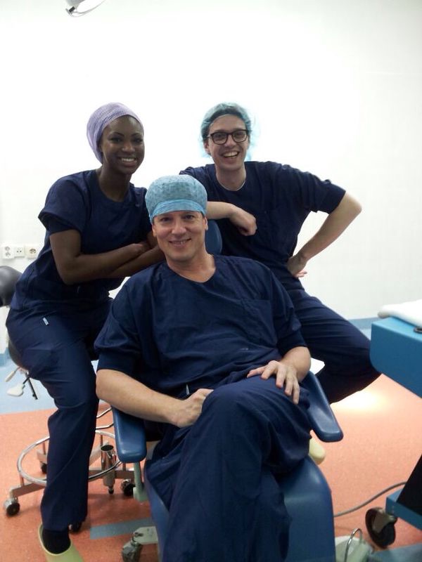
    Oculoplastic team in the operating room with senior surgeon Dr Dion Paridaens (foreground), Dr Dianta Pinas (left behind) and Dr Ronald de Keizer (right behind)

  • Over wallen onder de ogen en ooglid correctie

    Vermoeide uitdrukking

    In de focuskliniek van het Oogziekenhuis Rotterdam zie ik als cosmetisch ooglidchirurg veel cliënten met een verzoek voor een boven of onderooglid-correctie. De meesten hebben een milde tot matige mate van huidoverschot, sommige zelfs ernstig. Rokers presenteren zich vaak met festoons: een huidplooi, soms gevuld met vocht, dat als een soort zakje aan de onderkant van de onderoogleden hangt. Oogleden kunnen ook een vetoverschot vertonen, vooral opmerkelijk bij cliënten met overgewicht of bekende dermatochalazis (huidoverschot door verlies van elastinevezels). Wallen onder de ogen of huidoverschot in de bovenoogleden op basis van dermatochalazis dragen ​​bij aan een vermoeide uitdrukking, die vaak niet de energiestatus van de cliënt weergeeft.

    Ooglidcorrectie voor wallen onder de ogen versus bovenooglid correctie

    Blepharoplastieken van de onderoogleden (ooglidcorrectie) is vanuit chirurgisch oogpunt complexer en is minder vergevingsgezind dan de boven ooglidcorrectie, wat een zeer veel gebruikte procedure is. Beginnende chirurgen, vooral diegenen die door een fellowship-programma niet over de juiste oculoplastische chirurgische training beschikken, komen er al snel achter dat een operatie aan de onderoogleden nogal complex is, maar helaas ten koste gaat van een goed resultaat voor hun cliënten.

    Tijdens onderooglid correctie met huidverwijdering is canthopexy een essentiële stap: het ooglid wordt gefixeerd met een of twee (begraven) hechtingen aan de diepere weefsels in de laterale ooglidhoek om een postoperatieve verkeerde positie (ooglidretractie) te voorkomen die een onnatuurlijke en ongewenste uitstraling van oogwit onder de iris. (Copyright Dr. D Paridaens, oculoplastisch chirurg)

    Waarom is onderooglid correctie moeilijker dan bovenooglid correctie? Het belang van canthopexie of canthoplastiek bij ooglidcorrectie!

    Ten eerste zijn de incisies in de bovenste oogleden verborgen in de natuurlijk huidplooi (natural crease) van het bovenooglid. Slechts heel dicht bij de neus en aan de zijkant kunnen we een tijdelijk zichtbaar litteken zien, dat over het algemeen heel goed in de maand na de operatie heelt en in de beginfase gecamoufleerd kan worden met een foundation. In de onderoogleden kunnen de littekens gedurende weken tot maanden zichtbaar zijn, de laatste vooral bij rokers, ondanks het feit dat ze in de relaxed skin tension lines worden geplaatst (de natuurlijke huidlijnen die optreden tijdens het verouderingsproces) (hierdoor ontstaan als het ware “verborgen incisies”).

    Fixatie van het onderooglid aan het botvlies met begraven hechtingen en tevens diepe hechtingen buiten de laterale ooglidhoek geeft een goed en natuurlijk resultaat. Hierdoor wordt het onderooglid netjes om de oogbol heen gedrapeerd in een richting naar buiten en boven (zie pijl in figuur) en wordt de zwaartekracht geneutraliseerd. Als deze stappen niet worden uitgevoerd kan dit leiden tot postoperatieve complicaties: onjuiste positie en ontsierende retractie van de onderoogleden als ook droge ogen. (Copyright: Dr D Paridaens, oculoplastisch chirurg. Illustratie: Jessica Leenen)                                                                                                                                     

    Ten tweede kan er slapte van het onderooglid optreden als gevolg van verzwakking en veroudering van het ooglidweefsel en de ligamenten die het ooglid met het bot verbinden. Bovendien trekt de zwaartekracht de ooglidweefsels naar beneden. Als het laterale deel van het onderste ooglid niet voldoende wordt ingekort of gefixeerd aan het bot, kan een eventuele verwijdering van huidoverschot leiden tot een verkeerde positie van het ooglid (terugtrekking of retractie van het onderooglid), die nog kan worden versterkt door de zwaartekracht. Daarom moet de chirurg bij correctie van de onderoogleden dit trachten te voorkomen door middel van 1. adequate canthopexy of canthoplasty (procedures om het ooglid aan het bot te bevestigen) 2. beperking van de hoeveelheid verticale huidverwijdering tijdens blepharoplastieken, vooral wanneer het oog relatief uitstekend is of het jukbeen vlak is en 3. introductie van een ​​naar buiten / naar boven gerichte vector (krachtlijn) die de zwaartekrachtvector tegengaat door diep ondersteunende hechtingen in de zachte weefsels net buiten de laterale hoek van de oogleden te plaatsen.

    Loeb-Hamra techniek voor ooglidcorrectie: vet herverdeling naar de traangoot in plaats van verwijdering!

    Als er vet teveel bestaat in de onderoogleden terwijl de traangoot of de groeve tussen onderooglid en de wang diep is door weefselverlies (wat het effect van kringen om de ogen versterkt) is een vet herverdeling logischer dan het verwijderen ervan. Deze techniek van hergebruik van overtollig vet naar een plaats in de buurt met een tekort aan vet werd beschreven door Dr Loeb en ook door Dr Hamra. Hierbij wordt -na openen van het onderooglid via een opening onder de wimperlijn- een opening in het bindweefsel schot gemaakt waardoor het vet naar voren kan puilen. Vervolgens kan dit vet in een te maken pocket boven of onder het botvlies van de bovenkaak worden gebracht voor vulling van de traangoot. Zie onderstaande illustraties.

    Na het openen van het onderooglid wordt het bindweefselschot zichtbaar waarachter het vetteveel zich bevindt. Vlak bij de oogkastrand (arcus marginalia) wordt dit schot geopend om het vet naar voren te laten komen. (Copyright Dr Dion Paridaens, oculoplastisch chirurg. Illustratie: Jessica Leenen)
    Na het openen van het bindweefselschot (septum orbitale) kan het surplus aan vet dat de zwelling veroorzaakt door de opening worden getrokken en over de oogkas rand naar onderen worden gepositioneerd in een pocket boven (of onder) het botvlies. Hiermee wordt de holle groeve opgevuld als natuurlijke filler. (Copyright: Dr Dion Paridaens, oculoplastisch chirurg. Illustratie: Jessica Leenen)
    Het overtollige vetschortje kan over de onderste oogkas rand worden gedrapeerd in de richting die nodig is om de holle groeve onder het onderooglid op te vullen. (Copyright: Dr Dion Paridaens, oculoplastisch chirurg. Illustratie: Jessica Leenen)
    Links: de onderooglidhuid wordt ingesneden onder de wimperlijn. De huid en kringspierlaag wordt naar beneden getrokken zodat het bindweefselschot (septum, paars) en de vetzwelling hierachter zichtbaar wordt. Rechts: het septum wordt ter hoogte van de arcus marginalis geopend waarna het vet naar beneden gedrapeerd kan worden met afname van de ooglidzwelling tot gevolg. (Copyright: Dr Dion Paridaens, oculoplastisch chirurg. Illustratie: Jessica Leenen)
    Het vet dat teveel was in het onderooglid wordt gebruikt voor vulling van de groeve onder het onderooglid en gefixeerd aan de onderlaag met enkele of matrashechtingen. (Copyright: Dr Dion Paridaens, oculoplastisch chirurg. Illustratie: Jessica Leenen)
    Het vetoverschot wordt over de oogkas rand gedrapeerd. Hierdoor wordt de holle groeve opgevuld waardoor er minder relief optreedt in het onderooglid/wang complex met verbetering van de cosmetiek (Copyright: Dr Dion Paridaens, oculoplastisch chirurg. Illustratie: Jessica Leenen)

    Aanvullende behandeling na ooglidcorrectie met laser of trichloorazijnzuur (TCA peeling)

    Een goed uitgevoerde onderooglidcorrectie geeft in het algemeen een verbetering van de uitstraling doordat evidente zwellingen of huidoverschot verminderd zijn. Het is echter niet zo dat de huid weer even mooi zal worden als vroeger. Vaak zal er na een goed uitgevoerde correctie toch nog een geringe rimpeling of huidoverschot te zien zijn. Zou dit echter nog verdere gecorrigeerd worden door excisie (chirurgische verwijdering) dan bestaat het gevaar van onderooglid retractie, dat zowel cosmetische als functionele bezwaren kan geven. Als de huid rimpelig is kan wel worden overwogen om de huid te behandelen met laser of met trichloorazijnzuur (TCA peeling) voor een verbetering van het cosmetische beeld. Bij geringe afwijkingen kan in plaats van chirurgie ook gekozen worden voor primaire behandeling met laser of peeling.

    Reëele verwachtingen ten aanzien van operatieresultaat

    Het is zaak dat er met name ten aanzien van de t.o.v. bovenooglidcorrectie meer complexe onderooglidcorrectie reëele verwachtingen bestaan vooraf aan de ingreep om teleurstelling te voorkomen. In de praktijk zal het overgrote merendeel van goed voorgelichte en geselecteerde clienten die een goede onderooglidcorrectie ondergaan hebben heel tevreden zijn met het resultaat.


    Voor meer informatie kunt u een afspraak maken bij Dr Dion Paridaens, oculoplastisch chirurg in de Focuskliniek Oogziekenhuis Rotterdam. (klik op hyperlink)

    Voor functionele ingrepen kunt u zich laten verwijzen door de huisarts of specialist naar Dr Dion Paridaens in het Oogziekenhuis Rotterdam. (klik op hyperlink)

    Ervaren chirurg voor ooglidcorrectie

    Dr Paridaens heeft ruime ervaring opgebouwd in 27 jaar chirurgie: ruim 35.000 ingrepen werden verricht. Hij opereert 2 tot 4 dagen per week met een focus op oogleden, oogkas en traanwegen. Daarnaast is hij specialist op het gebied van botox behandelingen, waar hij al 23 jaar ervaring mee heeft. ‘Een natuurlijke uitstraling verkrijgen’ is altijd het motto

    Oculoplastisch team in het Dagcentrum met Dr Dion Paridaens (voorgrond), Drs Dianta Pinas (links achter) en Drs Ronald de Keizer (rechts achter)